What is Lipodermatosclerosis?
Lipodermatosclerosis is disease affecting the skin and the connective tissue and it causes a change of the lower legs. It often occurs in patients who have venous insufficiency. Lipodermatosclerosis is also known as hypodermitis scierodermiformis or sclerosing panniculitis. This condition is a type of lower extremity panniculitis (i.e. inflammation of subcutaneous fat). In this condition, the veins usually have difficulty in sending blood from the legs back to the heart.
Although the precise cause of lipodermatosclerosis is unknown, it is believed that venous insufficiency and obesity are a contributing factor.
Two thirds of the affected individuals are usually obese, and this condition can be identified when one’s legs resemble an inverted champagne bottle.
The disorder is more common in women with high body mass and a history of venous abnormalities.
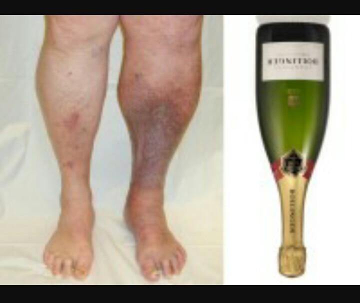
Fig.1 Legs resembling champagne bottle
Causes of Lipodermatosclerosis
Venous hypertension
Lipodermatosclerosis has been associated with venous hypertension- referring to raised pressure in the leg veins, which causes diffusion of substances such as fibrin, out of capillaries. When there is fibrotic tissues, this situation may result in ulceration.
A recurrent ulceration alongside fat necrosis are linked to lipodermatosclerosis. Venous hypertension can be as a result of standing upright or walking for long periods of time and unproductive calf muscle pump.
Venous incompetence
Venous incompetence (leaky valves) and obesity, relevant to underlying pathogenesis, can also cause the disorder.
Inflammation
Lipodermatosclerosis is also caused by an inflammation, which is caused by back pressure in the capillaries resulting in the activation of cells and soluble factors.
Coagulation
Changes in coagulation may also result into lipodermatosclerosis.
Two thirds of people with late fibrotic stage have obvious venous incompetence, venous hypertension, and extra-vacation of fluid from the veins, and end result is skin hardening and fibrosis.
Lipodermatosclerosis has originated from multifactorial issues involving tissue hypoxia, leaking of proteins right into the interstitium, as well as leukocyte activation. Studies have indicated a remarked decrease in concentration of oxygen, and this is linked to a decrease in capillary density.
In areas having fibrotic scars capillaries are almost absent, hence the condition known as livedoid vasculopathy or atrophie blanche.
Symptoms
Depending on the patient, this disorder may involve one or both of the lower inner legs. The disorder is characterized by skin induration or hardening, redness of the skin, increased pigment, swelling, and extensive fibrosis also referred to as sclerosis or scarring in the skin and subcutaneous tissue.
The patient may also experience tapering of the legs, especially above the ankles that forms a constricting band to give a shape like that of a champagne bottle placed upside down.
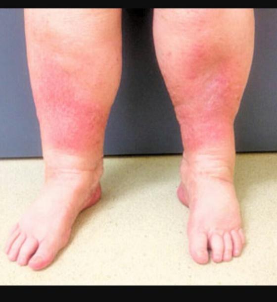
Fig.2 The condition can either be acute or chronic lipodermatosclerosis.
Acute and Chronic Lipodermatosclerosis
Lipodermatosclerosis can either be acute or chronic. With acute lipodermatosclerosis:
- Is not as a result of any other sickness or injury. A patient may experience painful inflammation, especially in the inner leg just above the ankle like the case of cellulitis.
- The affected area usually becomes red, tender, warm and may be scaly.
- The skin thickens but is not as sharp and painful as the chronic lipodermatosclerosis.
It should be noted that this largely affects the middle-age.
In the case of chronic lipodermatosclerosis it manifests in the following ways:
- Hardening of the skin and pain
- Localized thickening
- Moderate redness
- Varicose veins
- Increased pigmentation
- Small white scarred areas (atrophie blanche)
- Liquid released in the leg (oedema)
- Leg ulcers
The condition influences venous or stasis eczema.
Diagnosis
The disorder is usually diagnosed based on clinical and historical examinations.
Clinical examinations
Ultrasound scans and magnetic resonance imaging (MRI) tests may be used to define the extent to which the disease has affected the patient, or the best treatment approach or determine whether there is need for vascular surgery.
Skin biopsy or blood tests are not usually required to confirm diagnosis, but may be performed in only some rare cases.
Historical examinations
The doctor should be able to find out the history of previous skin problems and the relevant medical history, for example, lower leg injury, thrombosis, phlebitis and diabetes. A family history of varicose veins may increase the risk of chronic venous insufficiency.
It is also important to establish how the lower legs have changed in appearance or how any skin changes may have evolved.
Treatment
Compression therapy
The most common treatment is compression therapy to correct venous stasis or improve the venous insufficiency by reducing the edema. The compression stockings put enough pressure on the skin near the ankle while exerting less pressure on the patient’s calf.
Anabolic steroids
Anabolic steroids such as stanzolol, danzolol and oxandrolone have been used to enhance fibriosis and this helps with the cutaneous manifestations and painful nature of the lesions. The fibrinolytic agents use enzymatic action to help dissolve blood clots.
Weight reduction
One has to check weight and reduce excess weight as obesity has been associated with the condition. This calls for regular exercises.
Surgery
Doctors can also perform a vein surgery procedure, endovenous laser ablation or sclerotherapy and ultrasound therapy, which is also suitable to help patients with circulation.
Pentoxyfylline
A patient can use pentoxyfylline to reduce the increased blood flow and capsaicin to reduce pain.
Topical Steriod
Patients can receive clobetasel propionate, which is an ultrapotent topical steroid, or they may get intralesional triamcinolone injections to help in reducing inflammation.
Leg Elevation
One can also do a leg elevation, for example, raising legs on a pillow when you are resting or sleeping, not sitting or standing in one place for a long time, but when you do, bend and straighten legs every few minutes. This helps to keep blood moving in your legs back to the heart.
Keeping the skin well moisturized helps to keep the area around the ankles healthy. It is not however recommended to use topical antibiotics, such as neomycin, drying lotions such as calamine, lanolin, benzocaine or other creams that numb the skin.
Complications
Lipodermatosclerosis, ulceration, and venous disease may cause complications of immobility leading to increased body mass and other diseases associated with increased weight such as hypertension.
Support
There are some non-profit organizations that have come up to try support people living with this condition. These organizations bring awareness, provide patient information, and do research on better treatments and possible cures.
Pictures
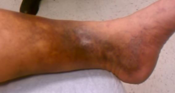
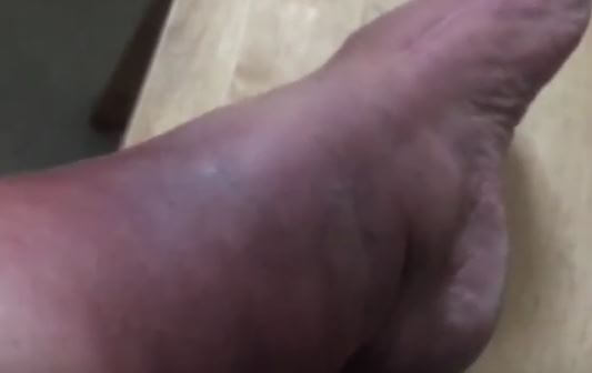
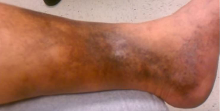
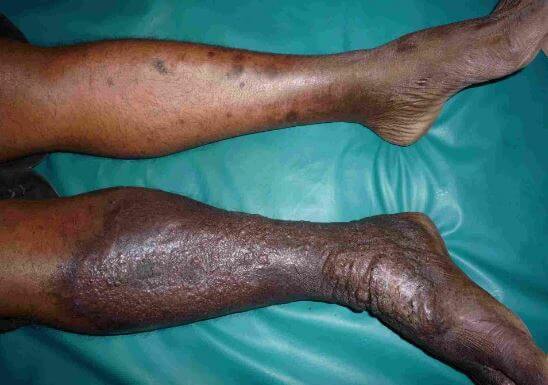
Image 6 – Acute Lipodermatosclerosis
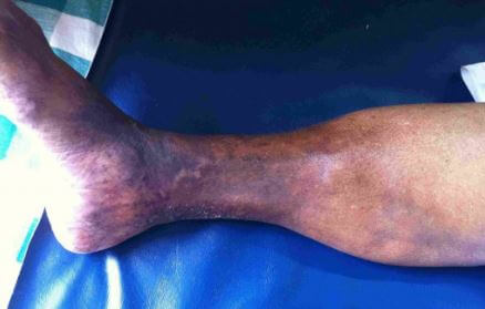
Image 7 – Chronic Lipodermatosclerosis
Reference List
- http://www.dermnetnz.org/topics/lipodermatosclerosis/
- https://en.wikipedia.org/wiki/Lipodermatosclerosis
- https://rarediseases.info.nih.gov/diseases/9671/lipodermatosclerosis
- Lipodermatosclerosis. Available at https://rarediseases.info.nih.gov/diseases/9671/lipodermatosclerosis/cases/21093
- Diagnosing Lipodermatosclerosis. Available at http://www.independentnurse.co.uk/clinical-article/diagnosing-lipodermatosclerosis/72537/
Very useful information, which will enable me to progress it with my GP.
I am no way a hypochondriac, simply interested in following up on an observation at Sunshine Coast University Hospital, Qld., with a very friendly and accommodating Doctor. I would anticipate that no follow procedures will be necessary or warranted.
Certainly not overweight – after a very energetic life, hormone therapy for Prostate Cancer stripped me of 25 Kg in no time flat, which has left me without energy and, at 73, little chance of regaining that lost muscle (which was stripped) to do anything meaningful (although I can now lift the (dense) mattress to make the bed which was not possible 2 years ago). I accumulated far too many ‘things-to-do’ prior!