What is a retracted eardrum?
Tympanic membrane retraction is a condition, where a part of the eardrum becomes weakened and loose and is pulled inwards by the negative pressure of the middle ear. The membrane can drop over the ossicles and cause a variety of symptoms. Retractions can vary in size- they can be small, with only a small area retracted in otherwise normal eardrum, or they can be very large and impair hearing. There are several names for retracted eardrum, but they all basically mean the same:
- Retraction
- Retraction pocket
- Atelectasis of the eardrum[1,4]
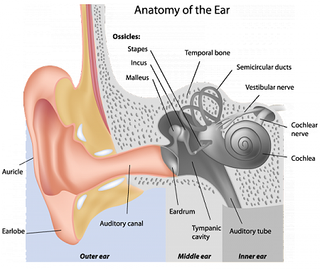
Anatomy of the eardrum
The eardrum (tympanic membrane) is a semi-transparent membrane that receives sound vibrations from outer air and transmits them to the ossicles in the middle ear. It also separates the external and internal auditory canal. The membrane is usually 9×11 mm in diameter. The eardrum is attached to the ring of bone-tympanic annulus. This part is called pars tensa, and it makes up ¾ of the eardrum. The central part of it is attached to the bone of inner ear, and it is more loose- pars flacida (¼ of the eardrum). The central part of the eardrum is pointed inward. The tympanic membrane is in 45-degree angle. Tympanic membrane has 3 layers:
- Outer layer is made of skin from the external auditory tube
- Middle part is made of two types of connective tissue fibers
- Inner part is covered with mucosa from the inner auditory tube [2].
Pathophysiology
Retracted eardrum develops if there is a long-term negative pressure from the inner part of tuba auditiva (Eustachian tube). The negative pressure in the middle ear pulls the eardrum inwards. This process happens more easily, if the eardrum is already weakened by a previous condition, like otitis media, ear surgery or trauma. Because of these conditions, the middle layer of eardrum can fade and make the eardrum weaker [4].
Types
J. Sade (1976) classified tympanic retraction into 5 grades:
- Mild tympanic membrane retraction
- Tympanic membrane retraction in contact with incus or stapes (ossicles in middle ear)
- Tympanic membrane in contact with promontory wall
- Tympanic membrane is adhered to promontory (adhesive otitis media)
- Grade III or IV with perforation of tympanic membrane
Another way to classify eardrum retraction is depending on the part which is retracted:
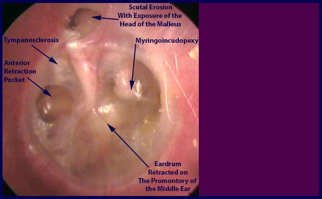
- Pars tensa retraction- more common and frequently seen in association with erosions of the ossicular bones, especially the long process of the incus
- Pars flaccida retraction- seen less commonly. This type of retraction is a predisposing factor for cholesteatomas [3,6].
![]()
Causes
Most common causes of eardrum retraction are:
- Middle ear infection (otitis media with effusion or serous otitis media)(Also see CHARGE syndrome)
- Auditory tube dysfunction
- History of eardrum rupture
- Eardrum ruptures chronically
- Ruptured eardrum does not heal properly [6]
- Ear infections are often seen in Velocardiofacial syndrome.
Symptoms
The symptoms of retracted eardrum vary depending on the severity of the condition. Most commonly the symptoms are:
- Pain in the ear
- Loss of hearing (conductive hearing loss)
- Acute suppurative otitis media
- Tinnitus- ringing in the ear
- Vibrating sensation in the ear
- Discharge from the ear
- Sensation of foreign object[5]

![]()
Diagnosis
The diagnosis mainly consists of thorough physical examination and imaging studies. Physical signs include:
- Poor movement of tympanic membrane
- Vascular dilatation
- The retracted area is translucent
- Tympanic membrane can be thickened
- Areas of indrawn eardrum
Other signs include worsening or loss of hearing in one ear, pain, discharge from the ear.
Best modality for imaging studies is magnetic resonance. MRI investigations should be done in preparation for surgery and afterwards. CT scanning can also be helpful in detecting possible causes for retraction, as well as for evaluating the progression of disease [4,5].
Treatment
Medications
Corticosteroids
Use of corticosteroids can change the course of retraction. They are useful in treating initial stages of retraction. The action of corticosteroids includes:
- Reduction of edema in Eustachian tube
- Reduction of ear secretions
- Reduction of allergic processes
Antibiotics
Antibiotics are used if there is an ongoing infectious process. If any bacterial infection is suspected, antibiotics should be used as adjuvant therapy. Middle ear infections are often seen in Edwards syndrome.
Other drugs
Releasing congestion can be helpful in treating retracted eardrum. Drugs like pseudoephedrine, anti-histamines, mucolytic drugs, nasal sprays can relieve symptoms of middle ear pathology [6].
Instrumental management
Instrumental management procedures are based on creating a pressure balance. Positive pressure exercises generate positive pressure input through the nose and Eustachian tube, which normalizes the pressure in tympanic cavity [6].
Surgical treatment
There are several procedures that can be done, but they depend on the progression of the disease.
Myringotomy
During myringotomy procedure, small incisions are made in tympanic membrane under local anesthesia. the secretions in the middle ear are than aspirated and the tympanic cavity is ventilated. This procedure is effective in milder cases (Grade I and sometimes Grade II).
Tympanostomy tubes
This procedure is the most frequently performed procedure in otorhinolaryngology. The tympanostomy tubes are placed in anterior and posterior-inferior quadrants of the eardrum, and they prevent the buildup of fluid in the middle ear. They are indicated in Grade I to Grade III retraction. This method of treatment is also used in Meniere’s syndrome.
![]()
Tympanectomy
Tympanectomy procedure consists of two parts. Firstly, the retracted part of the membrane is resected. The perforated area is left to heal and close spontaneously. The second part of the procedure consists of placing a tympanostomy tube in the remaining tympanic area, to avoid development of a new retraction. This procedure is indicated for Grade II or higher retraction.
Tympanochondroplasty
During this procedure, an artificial middle layer of the eardrum is placed that cannot be depressed by the negative pressure. Usually a piece of cartilage, harvested from the ear is used [6].
References
- What is retracted eardrum: http://www.entnet.org/content/conductive-hearing-loss-causes-and-treatments
- Anatomy of the ear: https://global.britannica.com/science/tympanic-membrane
- Types of retracted eardrum: https://radiopaedia.org/articles/tympanic-membrane-retraction
- Pathophysiology: http://www.allearseducation.org/resources/Tutorial-10-Retractions.pdf
- Symptoms: https://www.ncbi.nlm.nih.gov/pmc/articles/PMC4571482/
- Treatment: http://www.iapo.org.br/manuals/vi_manual_en_hector%20rondon.pdf
As Charlie Sheen says, this article is “WI!INNGN”